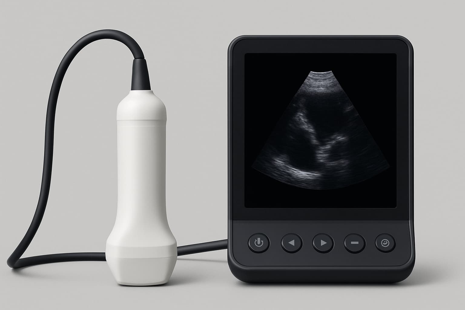
Point-of-care ultrasound is handheld cardiac imaging performed by the physician during the consultation itself. It allows structure and function to be assessed while the patient is in the room, answering many questions immediately that would otherwise require a separately scheduled study.
A New Era in Cardiac Imaging
In the past, evaluating heart health required bulky, expensive equipment and weeks of waiting for results. Today, thanks to advancements in handheld ultrasound technology — known as POCUS (Point-of-Care Ultrasound) — cardiologists can deliver immediate, high-quality imaging during the exam, not after.
What Is POCUS?
POCUS stands for "Point-of-Care Ultrasound." Unlike traditional ultrasound systems that are stationary and used by technicians in imaging departments, POCUS devices are portable, powerful, connected, and provide real-time results at the bedside or in the clinic.
Clinical Advantages of Handheld Ultrasound
- Rapid Diagnosis — Assess heart function, fluid buildup, valve abnormalities, or pericardial effusion within minutes
- Better Clinical Judgment — Real-time imaging enhances the physical exam
- Patient Engagement — Patients can see their own heart in real time
- Continuity of Care — Track changes over time with imaging at every visit
- Reduced Referral Delays — Many questions answered on the spot
POCUS empowers cardiologists to bring diagnostics directly to the patient, making imaging a seamless part of routine care.
The value is immediacy
A question raised during an examination can be answered during that examination rather than becoming a referral, a wait, and a second appointment to discuss results.
For a patient with a new symptom, that difference is often the difference between reassurance today and anxiety for three weeks.
It extends the physical examination
Bedside ultrasound does not replace listening and examining; it makes those findings more precise. A murmur can be looked at rather than only characterized by ear.
Used this way it is an extension of the clinical examination rather than a separate test.
Operator skill is the limiting factor
Image acquisition and interpretation both depend on training. The device has become inexpensive; competence has not.
It is reasonable to ask who is performing the study and what their training is, as with any imaging.
Knowing its limits is part of using it
A focused bedside study is not a complete echocardiogram and should not be presented as one. Subtle valve disease and detailed function assessment need the full study.
Used properly it triages: answering what it can, and identifying clearly what it cannot.
Documentation still matters
A bedside study should be recorded in the notes like any other examination finding, with images stored where they can be compared later.
Comparison over time is much of the value, and it is only possible if the earlier study was properly documented rather than glanced at and forgotten.
Common questions
- Is point-of-care ultrasound the same as a full echocardiogram?
- No. It is a focused bedside examination, not a replacement for a complete study. It answers specific questions quickly and identifies when a full echocardiogram is needed.
- Is it safe?
- Ultrasound uses sound waves rather than ionizing radiation, so there is no radiation exposure.
- Does it take extra time?
- It is performed during the visit and typically takes a few minutes, which is generally faster than arranging a separate appointment.
- What can it show?
- Cardiac chamber size and function, pericardial fluid, gross valve abnormalities, and volume status among other findings, depending on the clinical question.
- Will I still need other imaging?
- Sometimes. It frequently clarifies whether further imaging is needed, and what kind.